Venous Thromboembolism

Overview

Definition

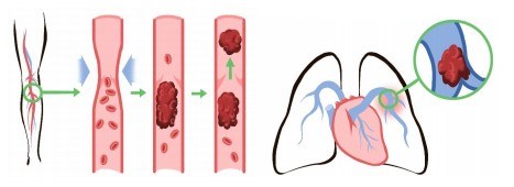
Venous thrombosis is the term used for a blood clot that forms within a vein. Venous thrombosis usually occurs below the knee in the deep calf veins and is most often associated with prolonged immobilization. Formation of venous thrombosis in the deep calf veins is referred to as a deep venous thrombosis or DVT.

Calf vein thrombosis can then propagate, in some cases into the lower thigh. If the clot breaks off and travels to the lungs, causing partial or full occlusion of pulmonary veins, it is called a pulmonary embolism (PE). 1 Together, DVT and PE are referred to as venous thromboembolism (VTE).

An illustration of a clot forming in the lower limb of an individual who has decreased blood flow (on the left) and an illustration of a clot which has broken off in a vein and resulted in a pulmonary embolus (on the right)

Prevalence

The reported incidence of DVT among patients with TBI ranges from 11% to 54%. 2-5 In the absence of prophylactic clot prevention, the estimated risk of developing DVT or PE is 20%. 6

Etiology

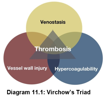
There are three primary risk factors for VTE, referred to as Virchow’s triad. Formation of VTE thrombi requires one or more of the factors in Virchow’s triad.

Virchow’ triad 7

  • Hypercoagulability
  • Venostasis (paralysis and immobility)
  • Vessel wall injury
  • Prolonged immobility is the factor most often associated with VTE and that is reflected in the risk factors listed below
  • Individuals with an ABI may enter a temporary trauma-induced hypercoagulable state 5,8
  • Endothelial or blood vessel inner wall damage contributes to the formation of thrombi in multiple ways

Risk Factors

  • Length of stay in the ICU
  • Number of days on a ventilator (>3days)
  • Paralysis
  • Immobility
  • Orthopedic surgery
  • Sepsis
  • Prior history of DVT
  • Advanced age
  • Congestive heart failure
  • Obesity
  • Myocardial infarction
  • Malignancy
  • Pregnancy
  • Oral contraceptive use
  • Smoking

Clinical Features

Deep Venous Thrombosis

  • Calf swelling but can involve whole leg swelling
  • Localized pain over deep veins
  • Redness, discoloration of skin in affected leg
  • Prominent superficial veins

Many DVT remain isolated to calf veins and are asymptomatic. Larger and more proximal DVTs are more likely to be symptomatic.

Pulmonary Embolus (PE)

  • Sudden shortness of breath
  • Increased heart rate
  • Chest pain
  • Pain with breathing (pleuritic pain)
  • Dizziness

PE can be small or large and again smaller pulmonary emboli may be asymptomatic while larger pulmonary emboli are more likely to be symptomatic.

Assessment

History

  • Clinical features listed above (note these clinical features overlap with a number of other medical conditions)
  • Recent immobility
  • Recent surgery
  • Lower extremity trauma
  • Presence of active cancer
  • Underlying chronic medical illness
  • Use of oral contraceptives
  • Family history of hypercoagulable state
  • History of spontaneous abortion (related to antiphospholipid antibody syndrome
  • Well’s criteria for PE or DVT (see screening)
  • PERC rule to rule out PE (see screening)

None of these criteria have been specifically verified in ABI patients.

Physical Exam

DVT

  • Examine for lower limb swelling (leg circumference 10 cm below the tibial tuberosity (> 3 cm difference between legs}
  • Focal tenderness over the path of deep veins (posterior calf, popliteal fossa and medial anterior thighs)
  • Color changes
  • Lower limb neurological exam (looking for paralysis)
  • Peripheral vascular exam

PE

  • Full cardiovascular exam
  • Full respiratory exam

OSCE Examination Guides

Screening

None of these screening tools have been specifically verified in ABI patients.

Well's Criteria for DVT

  • Active cancer
  • Bedridden recently >3 days or major surgery within 12 weeks
  • Calf swelling >3 cm compared to the other leg
  • Collateral (nonvaricose) superficial veins present
  • Entire leg swollen
  • Localized tenderness along the deep venous system
  • Pitting edema, confined to symptomatic leg
  • Paralysis, paresis, or resent plaster immobilization of the lower extremity
  • Previously documented DVT
  • Alternative diagnosis to DVT as likely or more likely

Well’s Score > 2 means DVT is likely

Well’s Score < 2 means DVT is unlikely

Well's Criteria for PE

  • Clinical signs and symptoms of DVT
  • PE is #1 diagnosis OR equally likely
  • Heart rate >100
  • Immobilization at least 3 days OR surgery in the previous 4 weeks
  • Previous, objectively diagnosed PE or DVT
  • Hemoptysis
  • Malignancy w/treatment within 6 months or palliative

Well’s Score > 4 means PE is likely

Well’s Score < 4 means PE is unlikely

PERC Rule (use to rule out PE)

  • Age ≥50
  • HR ≥100
  • O2 sat on room air <95%
  • Unilateral leg swelling
  • Hemoptysis
  • Recent surgery or trauma (surgery or trauma ≤4 weeks ago requiring treatment with general anesthesia)
  • Prior PE or DVT
  • Hormone use (oral contraceptives, hormone replacement or estrogenic hormones use in males or female patients)

If any criteria are positive, the PERC rule cannot be used to rule out PE in this patient.

Diagnostic Testing

Deep Venous Thrombosis

  • D-Dimer (breakdown product of cross-linked fibrin) – sensitive but non-specific for acute clot. If normal and Well’s Score is <2 than DVT is excluded; if elevated than move to ultrasound.
  • Venous duplex ultrasound 10 – easy and inexpensive – high sensitivity and specificity in acute symptomatic DVT – able to rule out DVT
  • Contrast venography – until recently considered gold standard for DVT but invasive and not recommended
  • Magnetic resonance venography 11 – less invasive than contrast venography but expensive and time consuming

Pulmonary Embolus

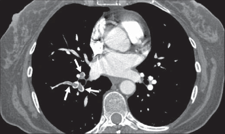
  • Computed tomography pulmonary angiogram 12
  • Pulmonary Ventilation (V) and Perfusion (Q) Scan 14
A pulmonary angiogram with arrows pointing to a pulmonary embolism. 13

Laboratory Investigations

DVT

  • D-dimer (see above)
  • Baseline testing:
    • INR
    • BUN/Cr
    • Liver function tests
    • CBC

PE

  • D-dimer (see above)
  • CK, Troponin (rule out cardiac ischemia)
  • Electrolytes
  • CBC (rule out infection)
  • Arterial blood gases (low blood oxygen levels – CO2 levels may also be low)

Diagnosis

Differential Diagnosis

DVT

  • Edema (often associated with paralysis of the lower extremity)
  • Heterotopic ossification
  • Fracture
  • Varicose veins
  • Superficial thrombophlebitis
  • Ruptured Baker cyst
  • Cellulitis
  • Lymphedema
  • Arterial insufficiency
  • Hematoma

PE

  • Pneumothorax
  • Myocardial infarction
  • Unstable angina
  • Pericarditis
  • Congestive heart failure
  • Pneumonia
  • Bronchitis
  • COPD
  • Asthma
  • Pleural effusion
  • Rib fracture
  • Muscular strain
  • Panic disorder

Complications

DVT

  • Post-phlebitic syndrome (chronic swelling of the involved leg due to damage to venous valves)
  • Pulmonary embolism

PE

  • Sudden cardiac death or heart failure
  • Obstructive shock
  • Atrial or ventricular arrhythmias
  • Secondary pulmonary arterial hypertension
  • Hypoxemia

Management

Management of VTE can be divided into: 1) Prevention and 2) Treatment.

Prevention

The incidence of VTE can be reduced in ABI

INESSS-ONF Clinical Practice Guideline Recommendations

Venous thromboprophylaxis should be initiated as soon as medically appropriate following traumatic brain injury. 25

Venous thromboprophylaxis should be initiated as soon as medically appropriate following traumatic brain injury. 15

  • Early frequent ambulation is recommended.
  • Pharmacological prophylaxis if high risk of DVT and low risk of bleeding – use low dose UFH or LMWH.
  • Mechanical, non-pharmacologic treatment if high risk DVT and high risk of bleeding – compression devices.

Treatment

  • Anticoagulation is the key to treatment.

Non-pharmacological Interventions

Prevention

Compression devices such as thromboembolism deterrent stockings, and intermittent pneumatic compression devices including arteriovenous foot pumps and sequential compression devices (SCDs) more for treatment of swelling.

  • They are not effective for prevention.
  • Overall, there is limited evidence in the ABI literature to support that compression devices are an effective prophylactic, particularly compared to low-molecular-weight heparin. 16-19
  • Multiple studies have found no significant differences between those who were treated with compression devices compared to those treated with pharmacological prophylaxis. 16,17
  • When pharmacological venous thromboprophylaxis is contraindicated or delayed after traumatic brain injury, physical methods (i.e., intermittent pneumatic compression stockings) should be utilized. 15
INESSS-ONF Clinical Practice Guideline Recommendations

When pharmacological venous thromboprophylaxis is contraindicated or delayed after traumatic brain injury, physical methods (i.e., intermittent pneumatic compression stockings) should be utilized. 25

InterventionEffectLevel of Evidence
Sequential Compression Devices-2
When compared to no sequential compression devices, sequential compression devices were no more effective in reducing the risk of DVT or PE

ERABI Evidence Table

Compression Devices Versus HeparinCC

Incidence of DVT or PE was no different between groups; however, mortality was higher in patients that received sequential compression devices alone


ERABI Evidence Table

Pharmacological Interventions

Prevention

InterventionEffectLevel of Evidence
Warfarin (Coumadin)+4
Associated with lower rates of DVT and PE, but increased risk of hemorrhagic bleeding

ERABI Evidence Table

Unfractionated Heparin-2
Within the first 72 hours, enoxaparin reduced the risk of DVT and PE compared to unfractionated heparin.

ERABI Evidence Table

Low-molecular-weight Heparin+4
Prophylactic enoxaparin (within the first 48h) reduces the risk of developing DVT and PE, without increasing the risk of bleeding in patients with severe ABI

ERABI Evidence Table

Enoxaparin Versus Unfractionated Heparin+2
Within the first 72 hours, enoxaparin reduced the risk of DVT and PE compared to unfractionated heparin.

ERABI Evidence Table

Treatment

DVT

Recommendations of the American College of Chest Physicians:

  • Proximal DVT of leg (above knee) – anticoagulation for a minimum of 3 months
  • Isolated DVT of leg (below knee) – high risk of extension or severe pain – treat with anticoagulation
  • Isolated DVT of leg – Acute isolated DVT without severe pain or high risk of extension – serial ultrasounds every 2 weeks

Anticoagulation with direct oral anticoagulants (dabigatran, rivaroxaban, apixaban) recommended over Warfarin which is in turn recommended over LMWH (enoxaparin).

Patients at high risk of bleeding (i.e., recent surgery) treat with intravenous VFH initially as it has a short half-life and can be reversed with protamine.

VTE and active cancer, obesity, pregnancy and hepatic disorders recommend LMWH

VTE with renal impairment treat with intravenous UFH followed by Warfarin.

Pulmonary Embolism

  • Treatment with anticoagulation

Surgical Interventions

Vena Cava Filters should be restricted to acute proximal DVT or pulmonary embolism where anticoagulation is not possible due to uncontrollable bleeding or high risk for life threatening bleeding or urgent surgery requiring anticoagulation be discontinued (Duffett et al. BMJ 2020; 370:m2177).

Once anticoagulation can be resumed IVC filter should be removed to limit complications.

InterventionEffectLevel of Evidence
Surgical EmbolectomyUCN/A

Although used clinically, surgical embolectomies have not been studied in a population with moderate to severe ABI.

Vena Cava FilterUCN/A

Although used clinically, vena cava filters have not been studied in a population with moderate to severe ABI.

Algorithm

Resources

References

1. Office of the Surgeon G, National Heart L, Blood I. Publications and Reports of the Surgeon General. In: The Surgeon General's Call to Action to Prevent Deep Vein Thrombosis and Pulmonary Embolism. Rockville (MD): Office of the Surgeon General (US); 2008.

2. Carlile M, Nicewander D, Yablon SA, et al. Prophylaxis for venous thromboembolism during rehabilitation for traumatic brain injury: A multicenter observational study. Journal of Trauma - Injury, Infection and Critical Care. 2010;68(4):916-923.

3. Cifu DX, Kaelin DL, Wall BE. Deep venous thrombosis: incidence on admission to a brain injury rehabilitation program. Arch Phys Med Rehabil. 1996;77(11):1182-1185.

4. Denson K, Morgan D, Cunningham R, et al. Incidence of venous thromboembolism in patients with traumatic brain injury. American journal of surgery. 2007;193(3):380-383; discussion 383-384.

5. Geerts WH, Code KI, Jay RM, Chen E, Szalai JP. A prospective study of venous thromboembolism after major trauma. The New England journal of medicine. 1994;331(24):1601-1606.

6. Haddad SH, Arabi YM. Critical care management of severe traumatic brain injury in adults. Scandinavian journal of trauma, resuscitation and emergency medicine. 2012;20:12.

7. Watanabe, Sant. Common medical complications of traumatic brain injury. . Physical Medicine and Rehabilitation: state of the art reviews. 2001;15:283-299.

8. Geerts WH, Jay RM, Code KI, et al. A comparison of low-dose heparin with low-molecular-weight heparin as prophylaxis against venous thromboembolism after major trauma. The New England journal of medicine. 1996;335(10):701-707.

9. Cifu DX, Caruso D. Traumatic brain injury. Demos Medical Publishing; 2010.

10. Pellecchia CM, Kory PD, Koenig SJ, et al. ACCURACY OF CRITICAL CARE PHYSICIANS IN THE ULTRASOUND DIAGNOSIS OF DEEP VENOUS THROMBOSIS (DVT) IN THE ICU. Chest. 2009;136(4):49S-49S.

11. Albert RK, Spiro SG, Jett JR. Clinical respiratory medicine. 3rd ed. Philadelphia: Mosby / Elsevier; 2008.

12. Fedullo PF, Tapson VF. Clinical practice. The evaluation of suspected pulmonary embolism. The New England journal of medicine U6 - ctx_ver=Z3988-2004&ctx_enc=info%3Aofi%2Fenc%3AUTF-8&rfr_id=info%3Asid%2Fsummonserialssolutionscom&rft_val_fmt=info%3Aofi%2Ffmt%3Akev%3Amtx%3Ajournal&rftgenre=article&rftatitle=Clinical+practice+The+evaluation+of+suspected+pulmonary+embolism&rftjtitle=The+New+England+journal+of+medicine&rftau=Fedullo%2C+Peter+F&rftau=Tapson%2C+Victor+F&rftdate=2003-09-25&rfteissn=1533-4406&rftvolume=349&rftissue=13&rftspage=1247&rft_id=info%3Apmid%2F14507950&rftexternalDocID=14507950¶mdict=en-US U7 - Journal Article. 2003;349(13):1247.

13. Stein EG, Haramati LB, Chamarthy M, Sprayregen S, Davitt MM, Freeman LM. Success of a Safe and Simple Algorithm to Reduce Use of CT Pulmonary Angiography in the Emergency Department. American Journal of Roentgenology. 2010;194(2):392-397.

14. Tran HA, Gibbs H, Merriman E, et al. New guidelines from the Thrombosis and Haemostasis Society of Australia and New Zealand for the diagnosis and management of venous thromboembolism. Medical Journal of Australia. 2019;210(5):227-235.
15. ONF-INESSS. Clinical Practice Guideline for the Rehabilitation of Adults with Modarate to Severe TBI. 2016.

16. Gersin K, Grindlinger GA, Lee V, Dennis RC, Wedel SK, Cachecho R. The efficacy of sequential compression devices in multiple trauma patients with severe head injury. The Journal of trauma. 1994;37(2):205-208.

17. Kurtoglu M, Yanar H, Bilsel Y, et al. Venous thromboembolism prophylaxis after head and spinal trauma: Intermittent pneumatic compression devices versus low molecular weight heparin. World Journal of Surgery. 2004;28(8):807-811.

18. Praeger AJ, Westbrook AJ, Nichol AD, et al. Deep vein thrombosis and pulmonary embolus in patients with traumatic brain injury: a prospective observational study. Critical care and resuscitation : journal of the Australasian Academy of Critical Care Medicine. 2012;14(1):10-13.

19. Minshall CT, Eriksson EA, Leon SM, Doben AR, McKinzie BP, Fakhry SM. Safety and efficacy of heparin or enoxaparin prophylaxis in blunt trauma patients with a head abbreviated injury severity score >2. The Journal of trauma. 2011;71(2):396-399; discussion 399-400.

20. Tran HA, Gibbs H, Merriman E, et al. New guidelines from the Thrombosis and Haemostasis Society of Australia and New Zealand for the diagnosis and management of venous thromboembolism. Medical Journal of Australia. 2019;210(5):227-235.