Neuroendocrine Disorders

Overview

Definition

Neuroendocrine Dysfunction

Neuroendocrine dysfunction is a common and potentially serious complication of acquired brain injury (ABI) that is increasingly recognized as a cause of morbidity in this population.

Neuroendocrine Disorders

Neuroendocrine disorders result from disruption of or injury along the hypothalamic-pituitary axis, an area of the brain that regulates physiological functions. 1

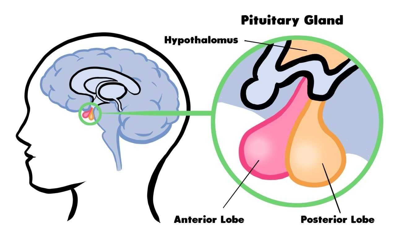
Anatomy of the pituitary gland


The following neuroendocrine disorders may occur as a result of acquired brain injury:

  • Syndrome of Inappropriate Antidiuretic Hormone Secretion (SIADH)
  • Diabetes Insipidus (DI)
  • Hyperprolactinemia
  • Hypoprolactinemia
  • Growth Hormone (GH) deficiency
  • Hypothyroidism
  • Adrenal insufficiency
  • Hypogonadism

Prevalence

The prevalence of neuroendocrine dysfunction varies widely across studies; estimates range from 20-70%. 2,3

  • The prevalence of specific hormone deficiencies has not been well-established in patients with ABI. This is due, in part, to variations in prevalence in the acute, subacute, and chronic phases of recovery.
  • Neuroendocrine disorders may be more likely to occur in the acute period after injury, while the long-term prevalence is likely closer to 30%.
  • The most common presentation of hypopituitarism following an ABI is a single-axis hormone deficiency, which is estimated to occur in 30-40% of patients who sustain an ABI compared to multi-hormone deficiencies, which are estimated to affect 10-15% of patients. 4-7
  • Of the single-axis hormone deficiencies, hyperprolactinemia (30%), GH hormone deficiency (30%), Diabetes insipidus (15-50%), adrenal insufficiency (9-80%), hypogonadism (10-30%), and hypothyroidism (10-30%) are the most well-described. 8-11

Etiology

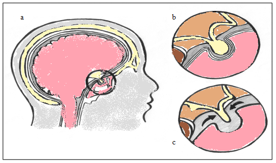
Most often associated with trauma to the pituitary gland in the area of the midbrain.

Pituitary Gland: Normal (b) and Post-Traumatic Conditions (c)
Hormonal DisorderPathophysiology
SIADH

- Excessive release of antidiuretic hormone from posterior pituitary

- Leads to hyponatremia and fluid overload

Central Diabetes Insipidus

- Decreased secretion of antidiuretic hormone

- Leads to hypernatremia
Hyperprolactinemia

- Results from a decrease in cerebral dopamine that causes an increase in prolactin release (i.e., prolactin is under tonic inhibition if dopamine)

Hypoprolactinemia

- Results from a decrease in prolactin

Growth Hormone Deficiency- Decreased growth hormone due to pituitary damage
Hypothyroidism- Decreased thyroid-stimulating hormone (TSH)
Adrenal Insufficiency- From a lack of corticotrophin releasing hormone or corticotropic hormone
Hypogonadism- Results from diminished production of follicle stimulating hormone (FSH) or luteinizing hormone (LH)

Risk Factors

  • Moderate to severe TBI
  • Injury severity
  • Glasgow Coma Scale 3-12
  • Location of Injury (basal skull fractures, diffuse axonal injury)
  • Increased intracranial pressure
  • Length of ICU stay

Clinical Features

Overall Clinical Features to look for:

  • Fatigue
  • Sleep disturbance
  • Decreased muscle mass, increased fat mass
  • Reduced exercise tolerance and muscle strength
  • Amenorrhea, decreased libido, erectile dysfunction
  • Decreased cognitive function, concentration, memory
  • Mood disturbances, depression, irritability
  • Social isolation, decreased quality of life

(note these clinical features overlap with non-endocrine conditions)

Hormonal AbnormalitiesClinical Features
SIADH
  • Often asymptomatic
  • Hyponatremia
  • May progress to confusion and hypoarousal
  • If severe enough can result in seizures (serum sodium <120)
Central Diabetes Insipidus
  • Hypernatremia
  • Polydipsia
  • Polyuria (>3L/24 hrs)
  • Excessive thirst
Hyperprolactinemia

Not clinically significant in men

In women:

  • Galactorrhea
  • Menstrual abnormalities
  • Hypogonadism
  • Sexual dysfunction
Hypoprolactinemia

In men:

  • Lactation
  • Breast enlargement
  • Decreased libido

In women:

  • Lactation
  • Breast enlargement or pain
  • Decreased libido
  • Oligomenorrhea or amenorrhea
Growth Hormone Deficiency
  • Fatigue
  • Exercise intolerance
  • Truncal obesity
  • Decrease in muscle mass
  • Confusion
Hypothyroidism
  • Fatigue
  • Cold intolerance
  • Dry skin
  • Peripheral edema
  • Constipation
  • Hair loss
Adrenal Insufficiency
  • Fatigue
  • Weakness
  • Weight loss
  • Hypoglycemia
  • Orthostatic hypotension
  • Anorexia
Hypogonadism

In men:

  • Infertility
  • Decreased libido
  • Pubic and body hair loss
  • Reduced facial hair growth
  • Erectile dysfunction

In women:

  • Infertility
  • Decreased libido
  • Pubic and body hair loss
  • Oligomenorrhea or amenorrhea

Assessment

History

  • Prior symptoms of endocrine dysfunction

Physical Exam

  • General medical examination
  • General neurologic examination
  • Cognitive testing

Screening

INESSS-ONF Clinical Practice Guideline Recommendations

Screening of the hypothalamic pituitary axis should occur at 3-6 months post traumatic brain injury or when symptoms are suggestive of a hormonal imbalance or deficiency. Screening should include a.m. cortisol, serum glucose, thyroid hormone (Free T4), thyroid-stimulating hormone (TSH), prolactin, estrogen or a.m. testosterone (T), follicle-stimulating hormone (FSH), luteinizing hormone (LH) and insulin-like growth factor-1 (IGF-1). Clinicians should be aware that a low or normal thyroid-stimulating hormone (TSH) does not rule out pituitary insufficiency with thyroid hormone deficiency. 13

Diagnostic Testing

Screening and Diagnostic Tests for Anterior Lobe Hypothalamic Pituitary Axis Dysfunction

Hormonal AbnormalitiesScreening TestsDiagnostic Testing
Hyperprolactinemia
  • Prolactin, estrogen or a.m. testosterone (T) – high consider medications
  • Screening tests enough for diagnosis; refer to endocrinologist
Hypoprolactinemia
  • Prolactin - low
  • Screening tests enough for diagnosis; refer to endocrinologist
Growth Hormone Deficiency
  • Insulin-like growth factor-1 (IGF-1) – low
  • Insulin-induced hypoglycemia test: cortisol <500 nmol/L
  • Insulin tolerance test: GH <5.1 ug/L
  • Glucagon stimulation test: GH <3 ug/L
Hypothyroidism
  • Thyroid hormone (free T4) - low


  • Thyroid-stimulating hormone (TSH) – low
  • Screening tests enough for diagnosis; refer to endocrinologist
Adrenal Insufficiency
  • a.m. cortisol (low is <7 ug/dL or 193 nmol/L)
  • Serum glucose (low is <4 without other explanation)
  • a.m. cortisol <100 nmol/L diagnostic
  • CRH stimulation test: cortisol <500 nmol/L

Screen and Diagnostic Tests for Posterior Lobe Dysfunction and Disorders of Sodium

Hormonal AbnormalitiesScreening TestsTesting
SIADH
  • Serum sodium <135 mmol/L
  • Urine sodium >40 mmol/L
  • Serum osmolality <280mmol/L
  • Urine osmolality >300 mmol/L
  • Clinical assessment and screening tests are sufficient for diagnosis.
  • Not dehydrated
  • If dehydrated likely Cerebral Salt Wasting
Central Diabetes Insipidus
  • Serum sodium >145 mmol/L
  • Urine sodium
  • Serum osmolality >290 mmol/L
  • Urine osmolality < 300 mmol/L
  • Urine output >3L/24h, or >200 mL/hr for 2+ consecutive hours
  • Clinical assessment and screening tests are sufficient for diagnosis.
  • Dehydrated

Distinguishing Disorders of Sodium Balance: DI, Cerebral Salt Wasting, and SIADH* (adapted from Rabinstein & Wijdicks 14)

Clinical or Lab FeatureDICSWSIADH
Serum sodiumHigh (>145mmol/L)Low (<135mmol/L)Low (<135mmol/L)
Urine sodiumVariableHigh (>20mmol/L)High (>20mmol/L)
Serum osmolalityHigh (>290mOsm/L)Low (<280mOsm/L)Low (<280mOsm/L)
Urine osmolalityLow (<300mOsm/L)High (>100mOsm/L)High (>300mOsm/L)
Total body volume (hydration status)DehydratedDehydratedNormal or excessive
Blood pressureLowNormal or lowNormal
HematocritHighHighNormal
Urine outputPolyuria (>3L of urine per 24hr, or >200 mL/hr for 2+ consecutive hrs)Polyuria (due to urinary loss of sodium)Normal or oliguria
Serum ADHLow (inappropriate)High (appropriate)

High (inappropriate)

*Laboratory values are provided for convenience based on normal reference values at St. Joseph’s Health Care London and London Health Sciences Centre. Please consult your lab’s normal values.

Laboratory Investigations

INESSS-ONF Clinical Practice Guideline Recommendations

Individuals with traumatic brain injury and hyponatremia should have an assessment of their hydration status, serum electrolytes with urinary electrolytes and sodium excretion. Restricting fluid intake and salt supplementation should be considered in managing the electrolyte disturbance in those with syndrome of inappropriate antidiuretic hormone secretion (SIADH) or hyponatremia due to cerebral salt wasting in individuals. 13

  • A.M. cortisol
  • Serum glucose
  • Thyroid hormone (free T4)
  • Thyroid-stimulating hormone (TSH)
  • Prolactin, estrogen or a.m. testosterone (T)
  • Follicle-stimulating hormone (FSH)
  • Luteinizing hormone (LH)
  • Insulin-like growth factor-1 (IGF-1)
  • Serum sodium and urine sodium
  • Serum osmolality and urine osmolality 15

Diagnosis

Differential Diagnosis

  • Other causes of endocrine dysfunction
  • Differential diagnosis is wide prior to screening because of overlapping clinical features with many other disease conditions

Management

Consult an Endocrinologist

Hormonal Abnormalities
SIADH
  • Fluid restriction of less than 1500 cc/day
  • Furosemide with sodium and potassium replacement
Central Diabetes Insipidus
  • Electrolyte abnormality (hypernatremia) can be life threatening
  • Treat dehydration with fluid replacement
  • Treat ADH deficiency with Desmopressin, an ADH replacement if necessary 16
Hyperprolactinemia
  • If do to anti-dopaminergic medications no treatment necessary
  • If not due to medications, consider treatment with dopamine agonist such as bromocriptine with referral to an Endocrinologist

Hypoprolactinemia

  • In women with fertility concerns refer to Endocrinologist
Growth Hormone Deficiency
  • Growth like Factor-1 Therapy
  • Recombinant GH 17-22
Hypothyroidism
  • Thyrotropin-Releasing Hormone or thyroid replacement
Adrenal Insufficiency
  • Hydrocortisone 23,24
Hypogonadism
  • Testosterone replacement in men
  • Estrogen replacement in women

Non-pharmacological Interventions

SAIDH

  • Fluid restriction

DI

  • Fluid volume

Pharmacological Interventions

InterventionEffectLevel of Evidence
Thyrotropin-Releasing Hormone (TRH)+4
TRH stimulation may be effective in treating hyponatremia

ERABI Evidence Table

Growth like Factor-1
Therapy
+1b
Growth hormone replacement therapy may improve clinical outcomes

ERABI Evidence Table

Growth like Factor-1
Therapy
+4
Growth hormone replacement therapy may be effective in treating GHD, fatigue, and depression post ABI.

ERABI Evidence Table

Progesterone+1b
Improves Glasgow Outcome and functional independence long-term, but results may not be observed short-term

ERABI Evidence Table

Resources

References

1. Sandel ME, Delmonico R, MJ K. Sexuality, reproduction, and neuroendocrine disorders following TBI. In: Zasler ND, Katz DI, RD. Z, eds. Brain Injury Medicine: Principles and Practices. New York, NY: Demos Medical Publishing; 2007:673-695.

2. Sirois G. Neuroendocrine problems in traumatic brain injury. In:2009.

3. Makulski DD, Taber KH, Chiou-Tan FY. Neuroimaging in posttraumatic hypopituitarism. Journal of Computer Assisted Tomography. 2008;32(2):324-328.

4. Aimaretti G, Ambrosio MR, Benvenga S, et al. Hypopituitarism and growth hormone deficiency (GHD) after traumatic brain injury (TBI). Growth hormone & IGF research : official journal of the Growth Hormone Research Society and the International IGF Research Society. 2004;14 Suppl A:S114-117.

5. Lieberman SA, Oberoi AL, Gilkison CR, Masel BE, Urban RJ. Prevalence of neuroendocrine dysfunction in patients recovering from traumatic brain injury. Journal of Clinical Endocrinology and Metabolism. 2001;86(6):2752-2756.

6. Kelly DF, Gaw Gonzalo IT, Cohan P, Berman N, Swerdloff R, Wang C. Hypopituitarism following traumatic brain injury and aneurysmal subarachnoid hemorrhage: A preliminary report. Journal of Neurosurgery. 2000;93(5):743-752.

7. Benvenga S, Campenní A, Ruggeri RM, Trimarchi F. Hypopituitarism secondary to head trauma. Journal of Clinical Endocrinology and Metabolism. 2000;85(4):1353-1361.

8. Hadjizacharia P, Beale EO, Inaba K, Chan LS, Demetriades D. Acute Diabetes Insipidus in Severe Head Injury: A Prospective Study. Journal of the American College of Surgeons. 2008;207(4):477-484.

9. Bondanelli M, De Marinis L, Ambrosio MR, et al. Occurrence of pituitary dysfunction following traumatic brain injury. Journal of Neurotrauma. 2004;21(6):685-696.

10. Hannon MJ, Crowley RK, Behan LA, et al. Acute glucocorticoid deficiency and diabetes insipidus are common after acute traumatic brain injury and predict mortality. The Journal of clinical endocrinology and metabolism. 2013;98(8):3229-3237.

11. Olivecrona Z, Dahlqvist P, Koskinen LO. Acute neuro-endocrine profile and prediction of outcome after severe brain injury. Scandinavian journal of trauma, resuscitation and emergency medicine. 2013;21:33.

12. Schneider HJ, Kreitschmann-Andermahr I, Ghigo E, Stalla GK, Agha A. Hypothalamopituitary dysfunction following traumatic brain injury and aneurysmal subarachnoid hemorrhage: A systematic review. Journal of the American Medical Association. 2007;298(12):1429-1438.

13. INESSS-ONF. Clinical Practice Guideline for the Rehabilitation of Adults with Moderate to Severe TBI. Ontario Neurotrauma Foundation. https://braininjuryguidelines.org/modtosevere/. Published 2015. Accessed2019.

14. Rabinstein AA, Wijdicks EF. Hyponatremia in critically ill neurological patients. Neurologist. 2003;9(6):290-300.

15. ONF-INESSS. Clinical Practice Guideline for the Rehabilitation of Adults with Modarate to Severe TBI. 2016.

16. Capatina C, Paluzzi A, Mitchell R, Karavitaki N. Diabetes Insipidus after Traumatic Brain Injury. J Clin Med. 2015;4(7):1448-1462.

17. Dubiel R, Callender L, Dunklin C, et al. Phase 2 randomized, placebo-controlled clinical trial of recombinant human growth hormone (rhGH) during rehabilitation from traumatic brain injury. Frontiers in Endocrinology. 2018;9 (SEP) (no pagination)(520).

18. Hatton J, Rapp RP, Kudsk KA, et al. Intravenous insulin-like growth factor-I (IGF-I) in moderate-to-severe head injury: A phase II safety and efficacy trial. Journal of Neurosurgery. 1997;86(5):779-786.

19. Mossberg KA, Durham WJ, Zgaljardic DJ, et al. Functional Changes after Recombinant Human Growth Hormone Replacement in Patients with Chronic Traumatic Brain Injury and Abnormal Growth Hormone Secretion. J Neurotrauma. 2017;34(4):845-852.

20. Gardner CJ, Mattsson AF, Daousi C, Korbonits M, Koltowska-Haggstrom M, Cuthbertson DJ. GH deficiency after traumatic brain injury: Improvement in quality of life with GH therapy: Analysis of the KIMS database. European Journal of Endocrinology. 2015;172(4):371-381.

21. Devesa J, Reimunde P, Devesa P, Barberá M, Arce V. Growth hormone (GH) and brain trauma. Hormones and Behavior. 2013;63(2):331-344.

22. Moreau OK, Yollin E, Merlen E, Daveluy W, Rousseaux M. Lasting pituitary hormone deficiency after traumatic brain injury. J Neurotrauma. 2012;29(1):81-89.

23. Tan CL, Alavi SA, Baldeweg SE, et al. The screening and management of pituitary dysfunction following traumatic brain injury in adults: British Neurotrauma Group guidance. J Neurol Neurosurg Psychiatry. 2017;88(11):971-981.

24. Grossman AB. Clinical Review#: The diagnosis and management of central hypoadrenalism. J Clin Endocrinol Metab. 2010;95(11):4855-4863.