Dysphagia & Aspiration

Overview

Definition

Dysphagia

  • Difficulty or discomfort with swallowing 1
  • Dysphagia is associated with tracheal penetration and aspiration

Tracheal Penetration

  • Entry of material into the airway past the epiglottis, but not below the true vocal cords

Aspiration

  • Entry of oral material into the airway below the level of the true vocal cords
  • Many individuals with dysphagia do not have aspiration, as the two are not synonymous

Silent Aspiration

  • Entry of material below the level of the true vocal cords, without cough or outward sign of difficulty 2
  • Normally, when material is aspirated, it elicits throat clearing and/or coughing
  • However, aspiration can occur without any outward sign of difficulty and this is referred to as silent aspiration

Prevalence

Dysphagia

  • Reported to range from 26% to 70% among patients entering rehabilitation post-ABI 3

Aspiration

  • Reported to range from 25% to 71% in patients with ABI 4-6

Silent Aspiration

  • Not well documented
  • Reported to range from 33% to 38% in patients with ABI 7,8

Etiology

Structural

  • Cerebral or brain stem lesion (neurogenic dysphagia)
  • Damage to cranial nerves involved in swallowing (CN IX and X)
  • Weakness or incoordination of the facial, oral, palatal or pharyngeal muscles

Behavioural and/or cognitive deficits

Risk Factors

  • Translaryngeal (endotracheal) intubation 9
  • Traumatic/urgent intubation10
  • Tracheostomy 9
  • Oropharyngeal spasticity or myoclonus10
  • Severity of brain injury 11
  • Duration of coma 7
  • Lower Glasgow Coma Score on admission (GCS 3-5) 9
  • Severity of CT scan findings 9
  • Duration of mechanical ventilation 9
  • Severe cognitive and cognition disorders 9
  • Physical damage to oral, pharyngeal, laryngeal, and esophageal structures 9
  • Oral and pharyngeal sensory difficulties 9

Clinical Features

  • Coughing before or after swallowing
  • Choking
  • Complaint of difficulty swallowing
  • Wet vocal quality
  • Pocketing of food in cheeks
  • Unexpected weight loss
  • Abnormal chest x-ray
  • Increased drooling
  • Delay in voluntary initiation of the swallow reflex
  • Increased body temperature around meal time
  • Avoidance or refusal to eat
  • Slow, effortful eating
  • Loss of food from mouth

Assessment

INESSS-ONF Clinical Practice Guideline Recommendations

Individuals with traumatic brain injury should be referred in a timely fashion to an appropriately trained and certified professional for a complete assessment of swallowing function when they present with one or more of the following risk factors for aspiration post-injury:

  • Presence of a tracheostomy
  • Poor cognitive functioning
  • Hypoactive gag reflex
  • Redsuced pharyngeal sensation
  • Brainstem involvement
  • Difficulty swallowing oral secretions
  • Coughing/throat clearing or wet/gurgly voice quality after swallowing water
  • Choking more than once while drinking 50ml of water
  • Weak voice and cough
  • Wet-hoarse voice quality
  • Recurrent lower respiratory infections
  • Unexplained low-grade fever or leukocytosis
  • Immunocompromised state

13

INESSS-ONF Clinical Practice Guideline Recommendations

Individuals with traumatic brain injury who are tracheotomized and/or ventilator-dependent should have an assessment by an appropriately-trained and certified professional to determine appropriateness for Passy Muir Valve placement or capping of trachea tube in preparation for swallowing assessment to optimize swallow function.13

History

  • Current swallowing and nutritional status
  • Review results from previous assessments of swallowing
  • Past medical history (e.g. other neurological disorder, respiratory disorder)
  • List of medications
  • The presence of risk factors (see above) should alert the clinician to carefully assess for dysphagia

Physical Exam

General

  • Level of consciousness
  • Cranial nerve assessment (in particular V, VII, IX, X, XII)
  • Observe voice quality
  • Chest auscultation
  • Palpate the neck (hyoid, thyroid, and cricoid cartilages) for structural abnormalities
  • Inspection of oral cavity (e.g. dentition, oral lesions, ability to manage own saliva)

Bedside oral motor assessment (usually done by a speech language pathologist)

  • Dry swallow – evaluate laryngeal elevation by palpation
  • Trial of 1-2 teaspoons of water
  • If well tolerated, trial a small cup of water
  • If water (a thin liquid) is not well tolerated, trial alternative liquid textures (see below)
  • Trial a cracker (regular dry food)
  • If the cracker is not well tolerated, trial alternative solid textures (see below)
  • Look inside oral cavity or use finger to sweep for food after swallow completed
  • Auscultate chest
  • Monitor oxygen saturation
  • Choking, coughing or wet gurgly voice are all suggestive of aspiration

*The presence or absence of aspiration cannot be reliably determined at the bedside

Screening

  • Screening is done at time of admission
  • May be done by trained nurse, OT or SLP
  • Involves assessing swallowing with a small amount of water (1-2 tablespoons or small cup) to see if patient chokes or coughs
  • Any sign of dysphagia patient remains NPO until full assessment

Diagnostic Testing

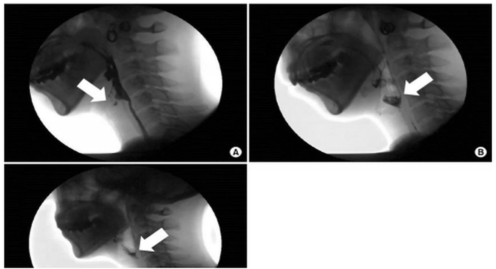
Example image of a VMBS assessment showing aspiration.15

Bedside swallowing evaluations are commonly used, however, there is limited evidence that they are effective

  • Only two clinical screening tests were found to be correlated with the findings on VMBS: 1) failure on 50 ml water test and 2) impaired pharyngeal sensation 14
  • There is limited evidence that clinical screening for dysphagia reduces pneumonia or length of hospital stay although it is considered the standard of care


Videofluoroscopic modified barium swallow (VMBS) is the gold standard

  • Involves radiation exposure
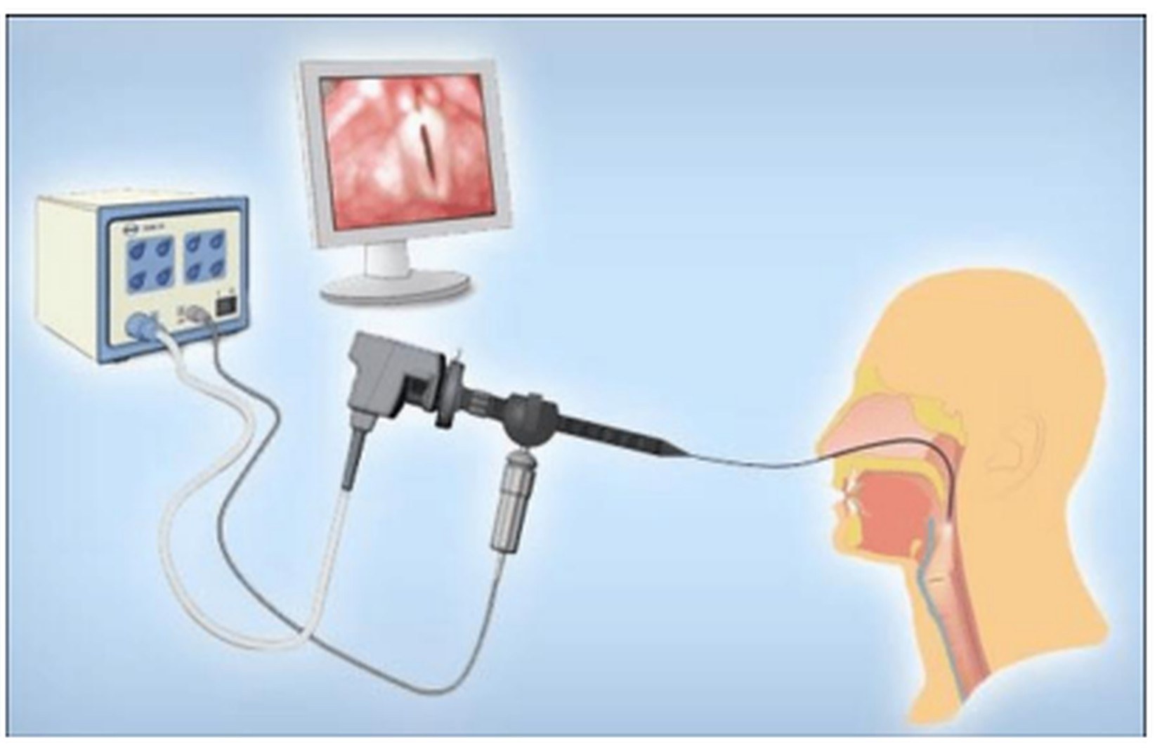
An illustration showing how FEES is administered.

Fiberoptic Endoscopic Evaluation of Swallowing (FEES)

  • Objective tool for the assessment of swallowing function and aspiration
  • Allows for the direct visualization of the pharynx before and after the swallow.
  • The camera view is obscured during the actual swallow and thus cannot show the mechanism underlying any observed dysfunction
  • Does not involve any radiation exposure
INESSS-ONF Clinical Practice Guideline Recommendations

For individuals with traumatic brain injury who are cognitively and physically able to tolerate it, videofluoroscopic modified barium swallow (VMBS) or modified barium swallow (MBS) studies should be used as a tool to assist in dysphagia management and identification of aspiration.13

INESSS-ONF Clinical Practice Guideline Recommendations

Instrumental assessment (VMBS) of dysphagia in post traumatic brain injury individuals should be considered when:

  • Effectiveness of compensatory strategies and techniques for safe swallowing is being evaluated
  • Bedside assessment indicates possible pharyngeal stage problems (which would potentially include the aspiration of food and fluid into the lungs)
  • The risks of proceeding on the basis of the bedside assessment outweigh the possible benefits (the person with TBI is at very high risk of choking or aspiration if fed orally)
  • The bedside assessment alone does not enable a sufficiently robust clinical evaluation to permit the development of an adequate plan for swallowing treatment

13

Laboratory Investigations

Blood work should be considered to assess nutrition and hydration status:

  • CBC
  • Electrolytes (sodium, potassium, chloride)
  • Extended electrolytes (calcium, magnesium, phosphate)
  • Renal function (urea, creatinine)

Diagnosis

  • Diagnosis begins early with screening for dysphagia followed by bedside assessment.
  • VMBS and FES are considered gold standards for objectively identifying presence, type and severity of dysphagia and aspiration.

Differential Diagnosis

  • Preexisting or posttraumatic structural abnormalities of esophagus and trachea
  • Psychogenic dysphagia (phagophobia)
  • Painful swallowing (odynophagia)
  • Globus pharyngis (“lump in one’s throat”)

Complications

  • Dysphagia can lead to malnutrition and dehydration
  • Aspiration can lead to serious complications including pneumonia, sepsis, and death

Management

INESSS-ONF Clinical Practice Guideline Recommendations

The dysphagia intervention plan for individuals with traumatic brain injury should incorporate an interdisciplinary approach and consider positioning, feeding strategies, medical status, pharmacological profile, cognitive impairments, behaviour, comfort and nutritional status.13

Non-pharmacological Interventions

Dietary Modifications

  • The evidence that dietary modifications affects the risk of aspiration pneumonia is not well established but is well accepted.
  • Generally, foods with a variety of consistencies are tried: Solids: puree, minced, chopped, soft and regular, Liquids: pudding, honey, nectar and thin.
  • The proper consistency of food or diet is dictated by VMBS studies and clinical assessment.
  • Thin liquids are typically harder to manage than thick fluids; thick fluids or even jelled water are used to eliminate thin liquids when this consistency proves hard to manage.
  • Regular solids are harder to manage than pureed diet.
  • Pureed diet with thick fluids is often used initially and can then progress to a dysphagia soft diet which eliminates all hard, small, and stringy food particles.
  • Sequential VMBS in complicated cases allows for progression of diet with swallowing recovery.

Low-Risk Feeding Strategies

  • It is important to encourage patients to feed themselves as the risk of aspiration pneumonia increases 20-fold when they are fed by someone else, generally because they are encouraged to eat at a faster rate.
  • Feed with hand-over-hand support at eye level if necessary.

Compensatory Strategies

  • There are a number of compensatory strategies for the ABI patient with dysphagia which are thought to reduce the risk of aspiration.
  • Patients should be fed in the upright posture.
  • Chin tuck facilitates forward motion of the larynx, thereby preventing food material from entering into the larynx and reducing the space between the base of the tongue and the posterior pharyngeal wall increasing pharyngeal pressure on the bolus moving through the pharynx.
  • Head rotation to the paretic side closes the ipsilateral pharynx, forces the bolus into the contralateral, less affected side of the pharynx and decreases cricopharyngeal pressures.
  • Head tilt uses gravity to guide the bolus into the ipsilateral pharynx.
  • Possible modified swallowing strategies may include:
    • Mendelsohn maneuver
    • Masako maneuver
  • Supraglottic swallow involves concomitant breath holding and swallowing which closes the tracheal vocal cords to protect the trachea. Supersupraglottic swallow adds the Valsalva maneuver to maximize vocal fold closing.
  • Double swallowing and coughing after swallowing helps to protect the airway.
  • Close supervision with cueing may be required to slow down impulsive fast eaters
  • Dysphagia therapy usually involves a combination of approaches, including exercises aimed at strengthening muscles, and improving movement and coordination.

Thermal Stimulation

  • For thermal stimulation, a cold stimulus is generally applied the anterior faucial pillar prior to the individual swallowing. This is believed to increase tactile and thermal stimuli, increasing oral awareness and can improve the transition from the initiated oral phase to the involuntary pharyngeal phase. 16

Modalities

  • Functional electrical stimulation may be used to enhance neuromuscular control of swallow
  • Neuromuscular electrical stimulation (NMES) focuses on peripheral stimulation of the oropharyngeal muscles to enhance neuroplasticity and recovery of swallowing function. 17
  • NMES results in greater muscular recovery than voluntary contraction due to recruiting a larger proportion of motor units. 18
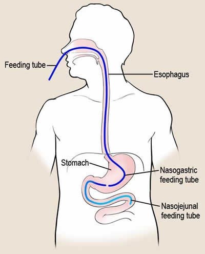
An illustration depicting a nasogastric tube.

Non-Oral Feedings

  • Non-oral feeding is a well-established practice for those patients who cannot handle oral feeds.
  • Non-oral feedings can be implemented almost immediately following a stroke in high risk patients using a naso-gastric tube.
  • If dysphagia is severe (i.e. patient is still aspirating in rehabilitation despite dietary modifications and compensatory strategies) and is expected to continue to do so for more than 6 weeks, a gastrostomy or jejunostomy tube is necessary.
  • Nasogastric (NG) tubes, usually intended for short-term use, are positioned directly into the stomach (with extensions into the small bowel) or small intestine through the nose and throat.
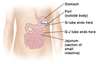
An illustration depicting a gastro-enteric tube.
  • Alternatively, gastro-enteric tubes are used for long-term feeding and are placed into the stomach percutaneously or surgically.

Prosthetics/Orthotics

  • Palatal augmentation prosthesis
  • Palatal lift prosthesis can elevate the soft palate, which improves bolus propulsion
  • One-way speaking valves in patient with tracheostomies allow for cough and subsequent cleaning of airway when aspiration occurs
InterventionEffectLevel of Evidence
Dietary ModificationsUCN/A

Although used clinically, dietary modifications have not been studied in a population with ABI.

Safe Eating Strategies UCN/A

Although used clinically, safe eating strategies have not been studied in a population with ABI.

Compensatory StrategiesUCN/A

Although used clinically, compensatory strategies have not been studied in a population with ABI.

Thermal/Tactile StimulationUCN/A

Although used clinically, thermal/tactile stimulation have not been studied in a population with ABI.

Oral Hygiene Education*+

2

Reduction of dental plaque, measured by the Plaque Index Score.

ERABI Evidence Table

Povidone Iodine+1b
Reduced incidence of ventilator-associated pneumonia.

ERABI Evidence Table

0.2% Chlorhexidine Gel

+1b
Reduced nosocomial infections and hospital length of stay.

ERABI Evidence Table

Oral Care*+1b
Reduced rates of pneumonia, febrile days, and pneumonia-related deaths.

ERABI Evidence Table

Non oral (enteral) feedingsUCN/A

Although used clinically, enteral feedings have not been studied in a population with ABI.

*Recommended by the INESSS-ONF Clinical Practice Guidelines

Pharmacological Interventions

There are no pharmacological interventions for the management of dysphagia and aspiration.

Surgical Interventions

  • If dysphagia is chronic and severe, a permanent tracheostomy with laryngectomy may be necessary to completely separate the airway from the food passageway
  • Pharyngeal bypass with gastrostomy to allow nutritional and hydration needs to be met through non oral feedings

Algorithm

Resources

References

1. Logemann JA. The evaluation and treatment of swallowing disorders. Current Opinion in Otolaryngology & Head and Neck Surgery. 1998.

2. Linden P, Siebens AA. Dysphagia: predicting laryngeal penetration. Archives of physical medicine and rehabilitation. 1983;64(6):281-284.

3. Ward E, Morgan, AT. Dysphagia following traumatic brain injury in adults and children: Assessment and characteristics. In: Murdoch BE, DG. T, eds. Traumatic brain injury: associated speech, language, and swallowing disorders. San Diego: Singular Publishing Group; 2001:331-367.

4. Mackay LE, Morgan AS, Bernstein BA. Factors affecting oral feeding with severe traumatic brain injury. The Journal of head trauma rehabilitation. 1999;14(5):435-447.

5. O'Neil-Pirozzi TM, Momose KJ, Mello J, et al. Feasibility of swallowing interventions for tracheostomized individuals with severely disordered consciousness following traumatic brain injury. Brain injury : [BI]. 2003;17(5):389-399.

6. Schurr MJ, Ebner KA, Maser AL, Sperling KB, Helgerson RB, Harms B. Formal swallowing evaluation and therapy after traumatic brain injury improves dysphagia outcomes. The Journal of trauma. 1999;46(5):817-821; discussion 821-813.

7. Lazarus C, Logemann JA. Swallowing disorders in closed head trauma patients. Archives of physical medicine and rehabilitation. 1987;68(2):79-84.

8. Terre R, Mearin F. Evolution of tracheal aspiration in severe traumatic brain injury-related oropharyngeal dysphagia: 1-year longitudinal follow-up study. Neurogastroenterology and motility : the official journal of the European Gastrointestinal Motility Society. 2009;21(4):361-369.

9. Mackay LE, Morgan AS, Bernstein BA. Swallowing disorders in severe brain injury: risk factors affecting return to oral intake. Archives of physical medicine and rehabilitation. 1999;80(4):365-371.

10. Cifu DX, Eapen BC, Janak J, Pugh M, Orman J. Traumatic Brain Injury Rehabilitation Medicine. Future Medicine; 2015.

11. Logemann JA. Evaluation and Treatment of Swallowing Problems. In: Zasler ND, Katz, D. I., Zafonte, R. D., ed. Brain Injury Medicine: Principles and Practice. 2 ed. New York: Demos Medical Publishing; 2013.

12. Horner J, Massey EW, Riski JE, Lathrop DL, Chase KN. Aspiration following stroke: clinical correlates and outcome. Neurology. 1988;38(9):1359-1362.

13. ONF-INESSS. Clinical Practice Guideline for the Rehabilitation of Adults with Modarate to Severe TBI. 2016.

14. Sørensen RT, Rasmussen RS, Overgaard K, Lerche A, Johansen AM, Lindhardt T. Dysphagia screening and intensified oral hygiene reduce pneumonia after stroke. Journal of Neuroscience Nursing. 2013;45(3):139-146.

15. Hun Lee S, Shin Lee E, Ho Yoon C, Shin H, Lee C. Collet-Sicard Syndrome With Hypoglossal Nerve Schwannoma: A Case Report. Vol 412017.

16. Malik SN, Khan MSG, Ehsaan F. Effectiveness of swallow maneuvers, thermal stimulation and combination both in treatment of patients with dysphagia using functional outcome swallowing scale. 2017.

17. Jayasekeran V, Singh S, Tyrrell P, et al. Adjunctive functional pharyngeal electrical stimulation reverses swallowing disability after brain lesions. Gastroenterology. 2010;138(5):1737-1746.

18. Sun SF, Hsu CW, Lin HS, et al. Combined Neuromuscular Electrical Stimulation (NMES) with Fiberoptic Endoscopic Evaluation of Swallowing (FEES) and Traditional Swallowing Rehabilitation in the Treatment of Stroke-Related Dysphagia. Dysphagia. 2013;28(4):557-566.